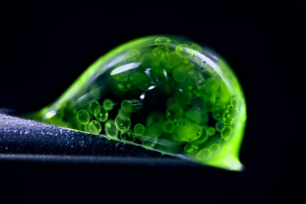
The Nikon Photomicrography Competition has once again showcased the captivating world of microscopy, revealing breathtaking images that challenge our perceptions of reality. This year, Jan Rosenboom, a chemical engineer from Germany, earned recognition as the runner-up with his stunning photograph of a droplet of water teeming with tiny spheres of colonial algae. The competition, which honors the remarkable contributions of microscopy to science, attracted numerous entries from both professional scientists and amateur enthusiasts.
Each year, the competition invites participants to submit images that capture the beauty and complexity of the microscopic world. This year’s collection features an array of subjects, including intricate human cellular networks and the fascinating hidden realms within ordinary fungi. The winners were announced on March 15, 2025, with numerous photographs highlighting the extraordinary details often overlooked by the naked eye.
Highlighting Nature’s Intricacies
Among the noteworthy entries was a breathtaking close-up of red pigments from the fungus Talaromyces purpureogenus, captured by Wim van Egmond from the Micropolitan Museum in the Netherlands. His image, which secured ninth place, illustrates the unexpected beauty that fungi can exhibit, showcasing their vibrant colors and complex structures.
Another significant contribution came from researchers at the Friedrich Miescher Institute for Biomedical Research in Switzerland, who utilized confocal microscopy to create a detailed image of a mouse colon. This technique, popular in biomedical research, enables scientists to study marked cells with fluorescent probes, providing insights into their structure and function.
The competition also featured an impressive image of heart muscle cells taken by James Hayes from Vanderbilt University. His photograph captures the dynamic processes of cell division, highlighting the intricate workings of our body’s cellular networks.
Wonders of the Microscopic World
One of the most striking images was created by Stella Whittaker from the National Institutes of Health (NIH). Her photograph, which resembles a swirling black hole, actually depicts induced pluripotent stem cell-derived sensory neurons. This image, achieved through a combination of advanced microscopy techniques, demonstrates how the microscopic world can defy initial appearances.
In a more sobering representation of nature’s complexity, Igor Robert Siwanowicz of the Howard Hughes Medical Institute illustrated the interdependent relationships within ecosystems. His photograph captures marrow pollen germinating on stigma while being parasitized by a filamentous fungus, showcasing the often brutal realities of survival in the microscopic realm.
The competition’s grand prize went to Zhang You, an entomologist from China, for his mesmerizing photograph of a rice weevil spreading its wings while perched on a single grain of rice. This exceptional image, which is a composite of over 100 stacked photographs, reflects Zhang’s dedication to ecological and insect science photography.
The Nikon Photomicrography Competition not only celebrates the artistry of microscopy but also emphasizes its vital role in scientific research. As these images reveal, the microscopic world is filled with beauty, complexity, and a myriad of life forms that continue to inspire and educate.